Google

Tuesday, July 31, 2007

Head and Face Reconstruction

Skull

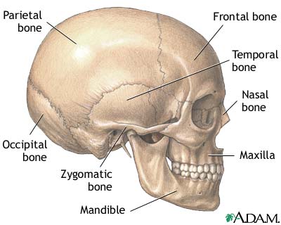
Skull

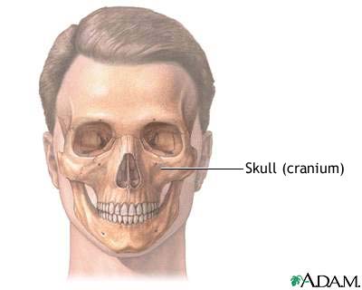
The skull is anterior to the spinal column and is the bony structure that encases the brain. Its purpose is to protect the brain and allow attachments for the facial muscles. The two regions of the skull are the cranial and facial region. The cranial portion is the part of the skull that directly houses the brain and the facial portion includes the rest of the bones of the skull.

Skull

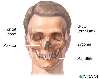
The skull is the bony structure of the head and face. The cranium surrounds the brain with the temporal, frontal, parietal and occipital bones. The maxilla, or upper jaw, and the mandible, or lower jaw, support the facial features of nose, mouth and eyes.

Cleft lip repair - series: Normal anatomy

Normal anatomy
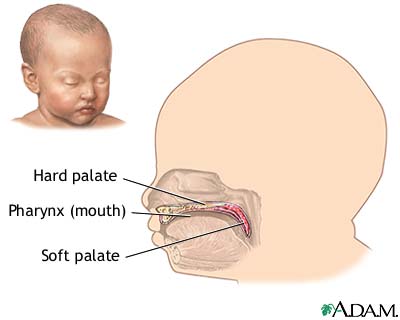
A cleft lip is an abnormal opening in the middle of the upper lip. A cleft palate is an opening in the roof of the mouth (palate).

Craniofacial reconstruction - series: Normal anatomy

Normal anatomy
There are four major bones of the face: the maxilla, the zygoma, the mandible, and the frontal bone of the cranium.

Definition

Surgical treatment to repair deformities of the head and face (craniofacial).

Description

Surgery for head and face deformities (craniofacial reconstruction) depends on the type and severity of deformity, and the condition of the patient.

In some cases, because surgical repairs involve the skull (cranium), brain, nerves, eyes, facial bones, and facial skin, a plastic surgeon (for skin and face) and a neurosurgeon (brain and nerves) work together. Head and neck surgeons may also perform craniofacial reconstruction operations.

The surgery is done while the patient is deep asleep and pain-free (under general anesthesia), and may take from 4 to over 12 hours to complete. Some of the facial bones are cut and repositioned into a more normal facial structure.

Pieces of bone (bone grafts) may be taken from the pelvis, ribs, or skull to fill in the spaces where bones of the face and head have been moved. Small metal screws and plates may be used to hold the bones in place. The jaws may be wired together to hold the new bone positions in place.

If the surgery is expected to cause much swelling of the face, mouth, or neck, the airway can become blocked. If this is anticipated, the patient may have what is called a tracheotomy placed, in which a small hole is made in the neck through which a tube (endotracheal tube) is placed in the airway (trachea).

This allows the patient to breath despite severe swelling of the face and upper airway that occurs after some operations and may persist for weeks.

Indications

Guidelines for craniofacial reconstruction include:

  • birth defects (such as hypertelorism, Crouzon's disease, Apert's syndrome)
  • injuries to the head, face, or jaws (maxillofacial)
  • tumors
  • deformities caused by treatments of tumors
Risks

Risks for any anesthesia are:

  • reactions to medications
  • problems breathing

Risks for any surgery are:

  • bleeding
  • infection

Additional risks of surgery of the head and face are:

  • nerve damage (cranial nerve dysfunction)
  • permanent scarring
  • partial or total loss of bone grafts


  • need for follow-up surgery

Saturday, July 14, 2007

Heart Attack

Acute MI

Acute MI

A heart attack or acute myocardial infarction (MI) occurs when one of the arteries that supplies the heart muscle becomes blocked. Blockage may be caused by spasm of the artery or by atherosclerosis with acute clot formation. The blockage results in damaged tissue and a permanent loss of contraction of this portion of the heart muscle.

Post myocardial infarction ECG wave tracings

Post myocardial infarction ECG wave tracings

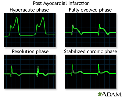
Various phases can be seen through ECG wave tracings following a heart attack:

  • Hyperacute phase begins immediately after a heart attack
  • Fully evolved phase starts a few hours to days after a heart attack
  • Resolution phase appears a few weeks after a heart attack
  • Stabilized chronic phase is the last phase and typically has permanent pathological changes compared to a normal ECG tracing

Progressive build-up of plaque in coronary artery

Progressive build-up of plaque in coronary artery

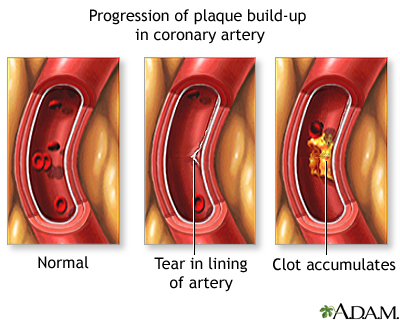
Plaque may build-up in a coronary artery at the site of a tear in the lining of the vessel.

Posterior heart arteries

Posterior heart arteries

The coronary arteries supply blood to the heart muscle. The right coronary artery supplies both the left and the right heart; the left coronary artery supplies the left heart.

Anterior heart arteries

Anterior heart arteries

The coronary arteries supply blood to the heart muscle. The right coronary artery supplies both the left and the right heart; the left coronary artery supplies the left heart.

Heart attack symptoms

Heart attack symptoms

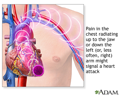
Symptoms of a possible heart attack include chest pain and pain that radiates down the shoulder and arm.


A heart attack is when low blood flow causes the heart to starve for oxygen. Heart muscle dies or becomes permanently damaged. Your doctor calls this a myocardial infarction.

Causes, incidence, and risk factors

Most heart attacks are caused by a blood clot that blocks one of the coronary arteries. The coronary arteries bring blood and oxygen to the heart. If the blood flow is blocked, the heart starves for oxygen and heart cells die.

A clot most often forms in a coronary artery that has become narrow because of the build-up of a substance called plaque along the artery walls. (See: atherosclerosis) Sometimes, the plaque cracks and triggers a blood clot to form.

Occasionally, sudden overwhelming stress can trigger a heart attack.

It is difficult to estimate exactly how common heart attacks are because as many as 200,000 to 300,000 people in the United States die each year before medical help is sought. It is estimated that approximately 1 million patients visit the hospital each year with a heart attack. About 1 out of every 5 deaths are due to a heart attack.

Risk factors for heart attack and coronary artery disease include:

Higher-than-normal levels of homocysteine, C-reactive protein, and fibrinogen may also increase your risk for a heart attack. Homocysteine is an amino acid. C-reactive protein and fibrinogen are linked to inflammation. Fibrinogen is also involved in blood clotting.

Symptoms

Chest pain is a major symptom of heart attack. However, some people may have little or no chest pain, especially the elderly and those with diabetes. This is called a silent heart attack.

The pain may be felt in only one part of the body or move from your chest to your arms, shoulder, neck, teeth, jaw, belly area, or back.

The pain can be severe or mild. It can feel like:

  • Squeezing or heavy pressure
  • A tight band around the chest
  • Something heavy sitting on your chest
  • Bad indigestion

Pain usually lasts longer than 20 minutes. Rest and a medicine called nitroglycerine do not completely relieve the pain of a heart attack.

Other symptoms of a heart attack include:

Signs and tests

A heart attack is a medical emergency. If you have symptoms of a heart attack, seek immediate medical help.

The health care provider will perform a physical exam and listen to your chest using a stethoscope. The doctor may hear abnormal sounds in your lungs (called crackles), a heart murmur, or other abnormal sounds.

You may have a rapid pulse. Blood pressure may be normal, high, or low.

Tests to look at your heart include:

Blood tests can help show if you have substances produced by heart tissue damage or a high risk for heart attack. These include:

Treatment

If you had a heart attack, you will need to stay in the hospital, possibly in the intensive care unit (ICU). You will be hooked up to an ECG machine, so the health care team can look at how your heart is beating. Life-threatening arrhythmias (irregular heart beats) are the leading cause of death in the first few hours of a heart attack.

The health care team will give you oxygen, even if your blood oxygen levels are normal. This is done so that your body tissues have easy access to oxygen, so your heart doesn't have to work as hard.

An intravenous line (IV) will be placed into one of your veins. Medicines and fluids pass through this IV. You may need a tube inserted into your bladder (urinary catheter) so that doctors can see how much fluid your body gets rid of.

THROMBOLYTIC THERAPY

Depending on the results of the ECG, certain patients may be given blood thinners within 12 hours of when they first felt the chest pain. This is called thrombolytic therapy. The medicine is first given through an IV. Blood thinners taken by mouth may be prescribed later to prevent clots from forming.

Thrombolytic therapy is not appropriate for people who have:

  • Bleeding inside their head (intracranial hemorrhage)
  • Brain abnormalities such as tumors or blood vessel malformations
  • Stroke within the past 3 months (or possibly longer)
  • Head injury within the past 3 months

Thrombolytic therapy is extremely dangerous in women who are pregnant or persons who have:

  • Severe high blood pressure
  • Had major surgery or a major injury within the past 3 weeks
  • Internal bleeding within the past 2-4 weeks
  • Peptic ulcer disease
  • A history of using blood thinners such as coumadin

MEDICINES FOR HEART ATTACKS

Many different medicines are used to treat and prevent heart attacks. Nitroglycein helps reduce chest pain. You may also receive strong medicines to relieve pain.

Antiplatelet medicines help prevent clot formation. Aspirin is an antiplatelet drug. Another one is clopidogrel (Plavix).

Beta-blockers (such as metoprolol, atenolol, and propranolol) help reduce the strain on the heart and lower blood pressure.

ACE inhibitors (such as ramipril, lisinopril, enalapril, or captopril) are used to prevent heart failure and lower blood pressure.

Wednesday, July 11, 2007

Heart-and-Lung Transplant

Lungs

Lungs

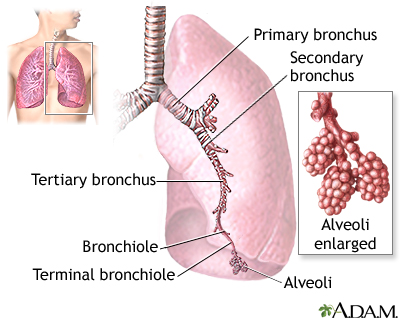
The major features of the lungs include the bronchi, the bronchioles and the alveoli. The alveoli are the microscopic blood vessel-lined sacks in which oxygen and carbon dioxide gas are exchanged.


Thoracic organs

Thoracic organs

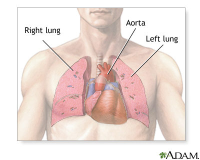
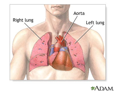
The thorax is also called the chest and contains the main organs of respiration and circulation. The heart through its main artery, the aorta, pumps oxygenated blood to all parts of the body. The lungs provide oxygen to the cells of the body and eliminate carbon dioxide. Together these organs sustain some of the most critical life functions of the body.


Heart-lung transplant - series: Normal anatomy

Normal anatomy
The heart and lungs are located in the thorax, or chest cavity. The heart pumps blood from the body to the lungs, where the blood is oxygenated. It then returns the blood to the heart, which pumps the freshly oxygenated blood to the rest of the body.
Heart-and-lung transplant is surgery to replace a diseased heart and lungs with a healthy heart and lungs from a human donor.

Description

Heart-and-lung transplant operations have been performed since 1980 in the United States. Since 1995, between 30 and 70 heart-lung transplants are performed each year, according to the United Network for Organ Sharing (UNOS).

The donated heart and lungs are from a person who has been declared brain-dead, but remains on life-support. Tissue matching helps find the best match of donated organs to the patient.

While the patient is deep asleep with general anesthesia, a cut is made through the breast bone. A heart-lung bypass machine takes over the circulating of the blood, and maintains oxygen levels to the body.

The patient's heart and lungs are removed, and the donor heart and lungs are stitched into place. The heart-lung bypass machine is disconnected. The blood flows through the donor heart and air flows in and out of the donor lungs.

Indications

Heart-lung transplant may be recommended for patients with:

Heart-and-lung transplants are not recommended for patients who have poor kidney or liver function, insulin-dependent diabetes mellitus, or other serious diseases.

Risks

Risks for any anesthesia are:

  • Reactions to medications
  • Problems breathing
Risks for any surgery are:
  • Bleeding
  • Infection
Additional risks of transplant includes:
  • Failure of the transplanted organs
  • Rejection of the transplanted organs
  • Infection due to anti-rejection (immunosuppression) medications
  • Blood clots
  • Stroke
Expectations after surgery

A heart-and-lung transplant extends the life of a patient who would otherwise die. The operation is done only when there is a very good chance of success. While long-term outcomes are unknown at this time, 5-year survival is about 40 - 50%.

As with all major organ transplants, the problems are finding a donor, preventing rejection, and the cost of the surgery and medications.

Finding a donor for heart-lung transplant can be difficult. The donated organs must come from a person who has been declared brain-dead, but is still on life-support. The patient who needs the transplant must be healthy enough to survive the surgery.

Preventing rejection is an ongoing process. The body's immune system considers the transplanted organs as invaders, and fights them.

To prevent rejection, organ transplant patients must take anti-rejection drugs such as cyclosporine and corticosteroids that reduce the body's immune response. These drugs also reduce the body's natural ability to fight off various infections.

Wednesday, July 4, 2007

Heart Failure

Heart, section through the middle

Heart, section through the middle

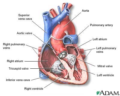
The interior of the heart is composed of valves, chambers, and associated vessels.

Heart, front view

Heart, front view

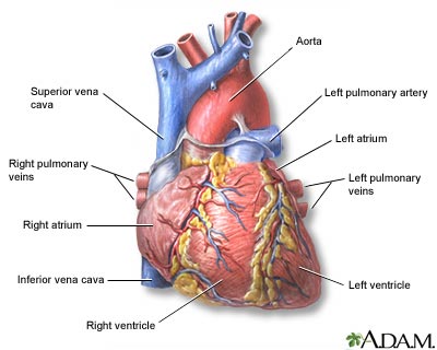
The external structures of the heart include the ventricles, atria, arteries and veins. Arteries carry blood away from the heart while veins carry blood into the heart. The vessels colored blue indicate the transport of blood with relatively low content of oxygen and high content of carbon dioxide. The vessels colored red indicate the transport of blood with relatively high content of oxygen and low content of carbon dioxide.

Circulation of blood through the heart

Circulation of blood through the heart

The heart is a large muscular organ which constantly pushes oxygen-rich blood to the brain and extremities and transports oxygen-poor blood from the brain and extremities to the lungs to gain oxygen. Blood comes into the right atrium from the body, moves into the right ventricle and is pushed into the pulmonary arteries in the lungs. After picking up oxygen, the blood travels back to the heart through the pulmonary veins into the left atrium, to the left ventricle and out to the body's tissues through the aorta.

Heart failure, also called congestive heart failure, is a life-threatening condition in which the heart can no longer pump enough blood to the rest of the body.

Causes, incidence, and risk factors

Heart failure is almost always a chronic, long-term condition, although it can sometimes develop suddenly. This condition may affect the right side, the left side, or both sides of the heart.

As the heart's pumping action is lost, blood may back up into other areas of the body, including:

With heart failure, many organs don't receive enough oxygen and nutrients, which damages them and reduces their ability to function properly. Most areas of the body can be affected when both sides of the heart fail.

The most common causes of heart failure are hypertension (high blood pressure) and coronary artery disease (for example, you have had a heart attack). Other structural or functional causes of heart failure include the following:

Heart failure becomes more common with advancing age. You are also at increased risk for developing heart failure if you are overweight, have diabetes, smoke cigarettes, abuse alcohol, or use cocaine.

Symptoms

Infants may sweat during feeding (or other exertion).

Some patients with heart failure have no symptoms. In these people, the symptoms may develop only with these conditions:

Signs and tests

A physical examination may reveal either an irregular or a rapid heartbeat. There may be distended neck veins, enlarged liver, swelling of the limbs (peripheral edema), and signs of fluid around the lungs (pleural effusion).

Listening to the chest with a stethoscope may reveal lung crackles or abnormal heart sounds. Blood pressure may be normal, high, or low.

An enlargement of the heart or decreased heart functioning may be seen on several tests, including the following:

This disease may also alter the following test results:

If excessive fluid has accumulated around the sac surrounding the heart (pericardium), you may need to have the fluid removed through a pericardiocentesis.

Treatment

If you have heart failure, your doctor will monitor you closely. This means having follow up appointments at least every 3 to 6 months, figuring out any underlying cause and treating it, and periodic testing of your heart function. For example, an ultrasound of your heart, called an echocardiogram, will be done once in awhile to give an estimate of how well your heart is pumping blood with each stroke or beat.

It is also your responsibility to carefully monitor yourself and help manage your condition. One important way to do this is to track your weight on a daily basis. Weight gain can be a sign that you are retaining fluid and that the pump function of your heart is worsening. Make sure you weigh yourself at the same time each day and on the same scale, with little to no clothes on.

Other important measures include:

  • Take your medications as directed. Carry a list of medications with you wherever you go.
  • Limit salt and sodium intake.
  • Don’t smoke.
  • Stay active. For example, walk or ride a stationary bicycle. Your doctor can provide a safe and effective exercise plan based on your degree of heart failure and how well you do on tests that check the strength and function of your heart. DO NOT exercise on days that your weight has gone up from fluid retention or you are not feeling well.
  • Lose weight if you are overweight.
  • Get enough rest, including after exercise, eating, or other activities. This allows your heart to rest as well. Keep your feet elevated to decrease swelling.

Here are some tips to lower your salt and sodium intake:

  • Look for foods that are labeled “low-sodium,” “sodium-free,” “no salt added,” or “unsalted.” Check the total sodium content on food labels. Be especially careful of canned, packaged, and frozen foods. A nutritionist can teach you how to understand these labels.
  • Don’t cook with salt or add salt to what you are eating. Try pepper, garlic, lemon, or other spices for flavor instead. Be careful of packaged spice blends as these often contain salt or salt products (like monosodium glutamate, MSG).
  • Avoid foods that are naturally high in sodium, like anchovies, meats (particularly cured meats, bacon, hot dogs, sausage, bologna, ham, and salami), nuts, olives, pickles, sauerkraut, soy and Worcestershire sauces, tomato and other vegetable juices, and cheese.
  • Take care when eating out. Stick to steamed, grilled, baked, boiled, and broiled foods with no added salt, sauce, or cheese.
  • Use oil and vinegar, rather than bottled dressings, on salads.
  • Eat fresh fruit or sorbet when having dessert.

Your doctor may consider prescribing the following medications:

  • ACE inhibitors such as captopril and enalapril -- these medications open up blood vessels and decrease the work load of the heart. These have become an important part of treatment
  • Diuretics -- there are several types including thiazide, loop diuretics, and potassium-sparing diuretics; they help rid your body of fluid and sodium.
  • Digitalis glycosides -- increase the ability of the heart muscle to contract properly; prevent heart rhythm disturbances
  • Angiotensin receptor blockers (ARBs) such as losartan and candesartan which, like ACE inhibitors, reduce the workload of the heart; this class of drug is especially important for those who cannot tolerate ACE inhibitors
  • Beta-blockers -- this is particularly useful for those with a history of coronary artery disease

Sometimes, hospitalization is required for acute CHF. Hospitalized patients may receive oxygen and intravenous medications such as vasodilators and diuretics. Medicines such as nesiritide (Natrecor) help dilate blood vessels and may also be helpful.

Medicines called inotropic agents help improve the heart's ability to pump blood. Such drugs include dobutamine and milrinone. They are given by IV.

Unstable patients receiving several medications usually need also hemodynamic monitoring with Swan-Ganz catheterization.

Severe cases of CHF require more drastic measures. For example, excess fluid can be removed through dialysis, and circulatory assistance can be provided by implanted devices such as the intra-aortic balloon pump (IABP) and the left ventricular assist device (LVAD). These devices can be life-saving, but they are not permanent solutions. Patients who become dependent on circulatory support will need a heart transplant.

A number of studies have shown that heart failure symptoms can be improved with a special type of pacemaker. It paces both the right and left sides of heart. This is referred to as biventricular pacing or cardiac resynchronization therapy. Ask your provider if you are a candidate for this.

Expectations (prognosis)

eart failure is a serious disorder that carries a reduced life expectancy. Many forms of heart failure can be controlled with medication, lifestyle change, and correction of any underlying disorder. Heart failure is usually a chronic illness, and it may worsen with infection or other physical stressors.

Complications

Possible side effects of medications include:

Prevention


Follow your health care provider's recommendations for treatment of conditions that may cause congestive heart failure. These recommendations may include:

  • Treat your high blood pressure with diet, exercise, and medication if necessary.
  • Treat your high cholesterol with diet, exercise, and medication if necessary.
  • DO NOT smoke
  • Avoid alcohol use
  • Take an ACE inhibitor if you have heart disease, diabetes, or high blood pressure.
  • Treat arrhythmias (abnormal heart rhythms) and keep your heart rate under control.
  • Treat an underlying thyroid disorder.

Also, consider the following lifestyle habits, especially if you have a strong family history of CHF:

  • Reduce salt intake.
  • Exercise

References

Hunt SA, Abraham WT, Chin MH, Feldman AM, Francis GS, Ganiats TG, et al. ACC/AHA 2005 Guideline Update for the Diagnosis and Management of Chronic Heart Failure in the Adult. J Am Coll Cardiol. 2005;46:1-82.
Hair Tonic

This poisoning is from swallowing hair tonic.

Poisonous Ingredient

Ethanol (ethyl alcohol)

Where Found

  • Various hair tonics

Symptoms

Home Treatment

DO NOT make a person throw up. Seek immediate medical help.

If the chemical was swallowed, immediately give the person water or milk, unless instructed otherwise by a health care provider. DO NOT give water or milk if the patient is having symptoms (such as vomiting, convulsions, or a decreased level of alertness) which make it hard to swallow.

If the chemical is in the eyes, flush with lots of water for at least 15 minutes.

Before Calling Emergency

Determine the following information:

  • The patient's age, weight, and condition
  • The name of the product (ingredients and strengths, if known)
  • The time it was swallowed
  • The amount swallowed

Monday, July 2, 2007

Hair Bleach Poisoning

This is poisoning from swallowing hair bleach.
Poisonous Ingredient
  • Hydrogen peroxide
  • Ethyl alcohol
  • Ammonium persulfate
Where Found
  • Some hair bleaches
  • Hydrogen peroxide
Note: This list may not be all inclusive.
Symptoms
Home Treatment

Seek immediate medical help.

If the chemical is on the skin or in the eyes, flush with lots of water for at least 15 minutes.

If the chemical was swallowed, immediately give the person water or milk, unless instructed otherwise by a health care provider. DO NOT give water or milk if the patient is having symptoms (such as vomiting, convulsions, or unconsciousness) that prevent swallowing.

Before Calling Emergency

Determine the following information:

  • The patient's age, weight, and condition
  • The name of the product (ingredients and strengths, if known)
  • The time it was swallowed
  • The amount swallowed


Immune Response

Immune system structures

Immune system structures

The immune system protects the body from potentially harmful substances. The inflammatory response (inflammation) is part of innate immunity. It occurs when tissues are injured by bacteria, trauma, toxins, heat or any other cause.

Phagocytosis

Phagocytosis

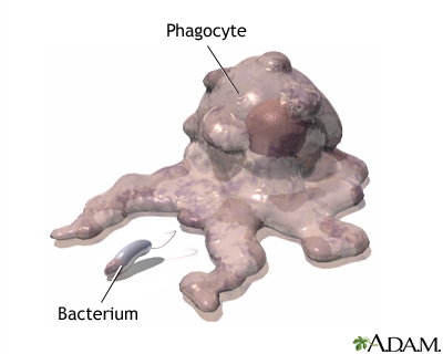
The chemicals also attract white blood cells that "eat" microorganisms and dead or damaged cells. The process where these white blood cells surround, engulf, and destroy foreign substances is called phagocytosis, and the cells are collectively referred to as phagocytes. Phagocytes eventually die. Pus is formed from a collection of dead tissue, dead bacteria, and live and dead phagocytes.

The immune response is how your body recognizes and defends itself against bacteria, viruses, and substances that appear foreign and harmful to the body.

The immune system protects the body from potentially harmful substances by recognizing and responding to so-called antigens. Antigens are large molecules (usually proteins) on the surface of cells, viruses, fungi, or bacteria. Non-living substances such as toxins, chemicals, drugs, and foreign particles (such as a splinter) can be antigens. Substances that contain these antigens are recognized and destroyed by the immune system. Even your own body cells have proteins that are antigens. These include a group of antigens called HLA antigens. Your immune system learns to see these antigens as normal and does not usually react against them.