Google

Wednesday, September 19, 2007

Urine - Bloody

Female urinary tract
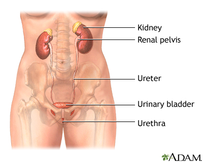
Female urinary tract

The female and male urinary tracts are relatively the same except for the length of the urethra.

Male urinary tract
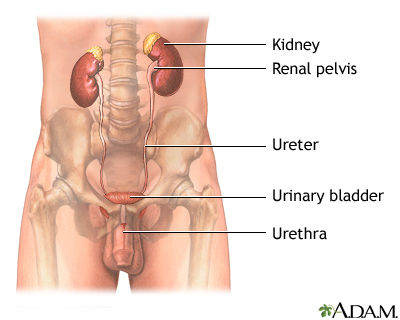
Male urinary tract

The male and female urinary tracts are relatively the same except for the length of the urethra.

Definition

Blood in your urine, or hematuria, can be classified as microscopic or gross.

  • Microscopic hematuria is when there is very little blood in the urine and it can only be seen with a microscope.
  • Gross hematuria is when there is enough blood in the urine that you can see it with the naked eye. Uusually it turns toilet water pale pink or bright red, or you may just see spots of blood in the water after urinating.

Considerations

In women, blood may appear to be in the urine when it is actually coming from the vagina. In men, what may be mistaken for urinary bleeding is sometimes a bloody ejaculation, usually due to a prostate problem.

Discoloration from certain drugs, beets, or other foods can mimic blood in the urine.

You may not see blood in your urine. In some cases, it is found microscopically when your doctor checks your urine during a routine exam. Your doctor will follow up on this problem to see if it persists and identify the cause.

When blood is visible to the naked eye, prompt and thorough evaluation is always needed. In children, hospitalization is often necessary to complete the work up.

Common Causes

There are many potential causes of blood in the urine. Often, bloody urine is from a problem in your kidneys or other parts of the urinary tract. If your kidneys, urinary tract, prostate, and genitals turn out to be fine, your doctor may check to see if you have a bleeding disorder.

Kidney and urinary tract causes include:

Causes from blood disorders include:

Call your health care provider if

Blood in the urine should never be ignored. Tell your doctor about this symptom and get an appropriate evaluation, especially if you have unexplained weight loss, burning with urination, frequent urination, or urgent urination.

Call your doctor right away if:

  • You have fever, nausea, vomiting, shaking chills, or pain in your abdomen, side, or back
  • You are unable to urinate
  • You are passing blood clots

Also call your doctor if:

  • You have pain with sexual intercourse or heavy menstrual bleeding -- the problem may be related to your reproductive organs
  • You have urine dribbling, nighttime urination, or difficulty starting your urinary flow -- the problem may be related to your prostate

What to expect at your health care provider's office

Your doctor will take a medical history and perform a physical examination. Medical history questions may include:

  • When did you first notice blood in your urine?
  • What is the underlying color of your urine?
  • Do you have any pain with urination?
  • Has the quantity of your urine increased or decreased?
  • Does your urine have an odor?
  • Are you urinating more frequently?
  • Do you have an urgent need to urinate?
  • What medications are you taking, including over the counter drugs?
  • Have you recently eaten foods that may cause discoloration, like beets, berries, or rhubarb?
  • Do you have any other symptoms like pain in your back, abdomen, or side? Fever, weight loss, nausea, vomiting, or diarrhea? Nighttime urination? Dribbling? Discharge from penis or vagina? Pain with intercourse?
  • Have you had previous urinary problems or kidney problems?
  • Do you have any allergies?
  • Have you had a recent injury?
  • Have you had any recent diagnostic or surgical procedures involving the urinary tract?

Tests that may be done include:

The treatment will depend on the cause of the blood in the urine. If a urinary tract infection is confirmed, antibiotics may be prescribed. If appropriate, pain medications will be given.
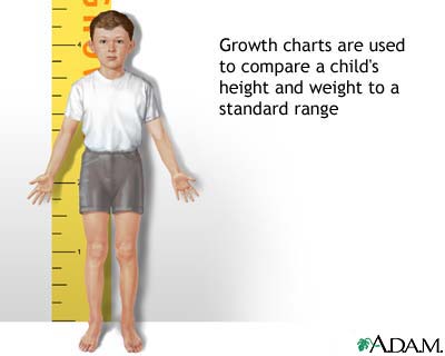
Height and Weight Chart

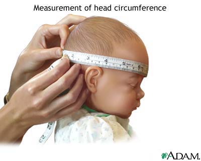
Head circumference

Head circumference

Head circumference is a measurement of the circumference of the child's head at its largest area (above the eyebrows and ears and around the back of the head). During routine check-ups, the distance is measured and compared to previous measurements. Normal ranges are based on sex and age.

Height/weight chart

Height/weight chart

A chart may be used to measure and compare a child's growth to a standard range. The parameters to be measured and charted are height, weight, and head circumference.

Information

A growth chart is used to measure and compare a child's growth with what is considered normal for that child's age and gender. The nationally accepted growth charts are based on measurement data collected by the National Center for Health Statistics. The parameters measured include height, weight, and head circumference.

Height and weight measurements are used to document a child's height or length (in inches or centimeters), weight (in ounces and pounds, or grams and kilograms), based on his age in weeks, months, and years. For children under 3 years, height is measured while they are lying down. For children over 3, height is measured while standing. Height and weight measurements are recorded and graphed until the child is 17.

Head circumference is a measurement of the size of the head taken by wrapping a tape measure above the eyebrows and around to the back of head.

Measurements are compared to the standard (normal) range for children of the same gender and age. Results are read as percentiles of average. For example, a child with a height at the 75th percentile means that only about 25% of children the same age and gender are taller and about 75% of children are shorter.

Growth charts are important in that they may provide an early warning that the child has a medical problem. For instance, during the first 18 months of life and particularly during early infancy, abnormal growth of the head can alert the doctor to a problem.

Growth that is too rapid may indicate the presence of hydrocephalus (water around the brain), a brain tumor, or other conditions that cause macrocephaly (abnormally large head). Growth that is too slow may indicate problems in brain development, early fusion of sutures (the bones of the skull), or other problems.

Insufficient gain in weight, height or a combination may indicate failure-to-thrive, chronic illness, neglect, or other problems.

Abnormal growth as seen on the growth charts is only an indicator of a potential problem. Your doctor will determine if it represents an actual medical problem or simply needs to be followed carefully. Because of individual variations in genetics and hormones, growth charts are not an accurate predictor of a child's future, full-grown height.

Saturday, September 15, 2007

Diabetes Diet

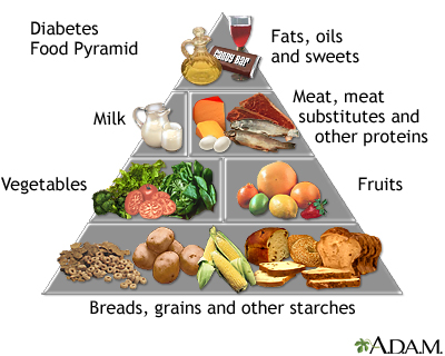
Diabetes Food Pyramid

Diabetes Food Pyramid

The Diabetes Food Pyramid divides food into six groups, which vary in size to show relative amounts of servings for each group.

This pyramid differs from the Food Guide Pyramid released by the USDA. In the Diabetes Food Pyramid, the groups are based on protein content and carbohydrates instead of their food classification.

Definition

Specific diabetic dietary guidelines have been developed by the American Diabetes Association and the American Dietetic Association to improve the management of diabetes.

Key principles are to:

  • Achieve weight control through reducing calories
  • Reduce intake of dietary fat (specifically saturated fat)
  • Individualize guidelines for carbohydrates based on the type of diabetes you have and the control of your blood sugar levels.

Function

There are 2 primary types of diabetes. The nutritional goals for each are different.

With type 1 diabetes, studies show that total carbohydrate has the most effect on the amount of insulin needed and the maintenance of blood sugar control. There is a delicate balance of carbohydrate intake, insulin, and physical activity that is necessary for the best blood levels of a sugar called glucose. If these components are not in balance, there can be wide fluctuations -- from too-high to too-low -- in blood glucose levels. For those with type 1 diabetes on a fixed dose of insulin, the carbohydrate content of meals and snacks should be consistent from day to day.

For children with type 1 diabetes, weight and growth patterns are a useful way to determine if the child's intake is adequate. Try not to withhold food or give food when a child is not hungry. Insulin dosing and scheduling should be based on a child's usual eating and exercise habits.

With type 2 diabetes, the main focus is on weight control, because 80% - 90% of people with this disease are overweight. A meal plan, with reduced calories, even distribution of carbohydrates, and replacement of some carbohydrate with healthier monounsaturated fats helps improve blood glucose levels.

Examples of foods high in monounsaturated fat include peanut or almond butter, almonds, walnuts, and other nuts. These can be substituted for carbohydrates, but portions should be small because these foods are high in calories.

In many cases, moderate weight loss and increased physical activity can control type 2 diabetes. Some people will need to take oral medications or insulin in addition to lifestyle changes.

Children with type 2 diabetes present special challenges. Meal plans should be recalculated often to account for the child's change in calorie requirements due to growth. Three smaller meals and 3 snacks are often required to meet calorie needs.

Changes in eating habits and increased physical activity help reduce insulin resistance and improve blood sugar control. When at parties or during holidays, your child may still eat sugar-containing foods, but have fewer carbohydrates on that day. For example, if birthday cake, Halloween candy, or other sweets are eaten, the usual daily amount of potatoes, pasta, or rice should be eliminated. This substitution helps keep calories and carbohydrates in better balance.

For children with either type of diabetes, special occasions (like birthdays or Halloween) require additional planning because of the extra sweets.

Recommendations

Reduce the amount of dietary fat. The current American Diabetes association guidelines advise that less than 7 - 10% of calories should come from saturated fat. These are the fats that raise LDL ("bad") cholesterol. Dietary cholesterol should be less than 200 - 300 mg per day. Additionally, intake of trans-unsaturated fats should be minimized. These are better known as partially hydrogenated oils. Reducing fat intake may help contribute to modest weight loss.

Keep protein intake in the range of 15 - 20% of total calories. Choices low in fat are recommended such as nonfat dairy products, legumes, skinless poultry, fish and lean meats. To keep the cholesterol content in range, approximately 6 ounces of protein per day is recommended. A portion of poultry, fish, or lean meat is about the size of 2 decks of cards.

Carbohydrate choices should come from whole grains breads or cereals, pasta, brown rice, beans, fruits and vegetables. Increasing dietary fiber is a general guideline for the entire population rather than specifically for people with diabetes. Portions and type of carbohydrate affect calories and is reflected by weight and blood glucose control. Learning to read labels for total carbohydrate rather than sugar provides the best information for blood sugar control.

Limit sources of high-calorie and low-nutritional-value foods, including those with a high content of sugars. Sugar-containing foods should be substituted for other carbohydrate sources (such as potatoes) instead of just adding them on to the meal.

A registered dietitian can help you best decide how to balance your diet with carbohydrates, protein and fat.

Friday, September 14, 2007

Diabetes

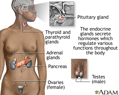
Endocrine glands

Endocrine glands

Endocrine glands release hormones (chemical messengers) into the bloodstream to be transported to various organs and tissues throughout the body. For instance, the pancreas secretes insulin, which allows the body to regulate levels of sugar in the blood. The thyroid gets instructions from the pituitary to secrete hormones which determine the pace of chemical activity in the body (the more hormone in the bloodstream, the faster the chemical activity; the less hormone, the slower the activity).

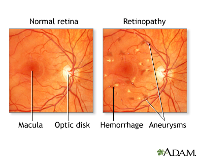
Diabetic retinopathy

Diabetic retinopathy

Diabetes causes an excessive amount of glucose to remain in the blood stream which may cause damage to the blood vessels. Within the eye the damaged vessels may leak blood and fluid into the surrounding tissues and cause vision problems.

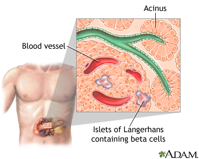
Islets of Langerhans

Islets of Langerhans

Islets of Langerhans contain beta cells and are located within the pancreas. Beta cells produce insulin which is needed to metabolize glucose within the body.

Blood test

Blood test

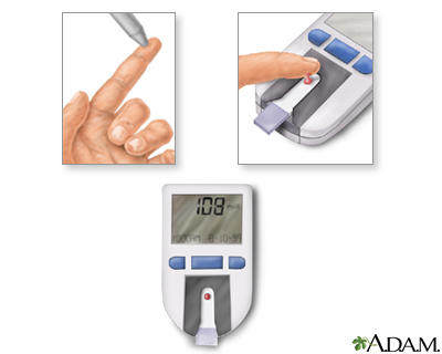
To monitor the amount of glucose within the blood a person with diabetes should test their blood regularly. The procedure is quite simple and can often be done at home.

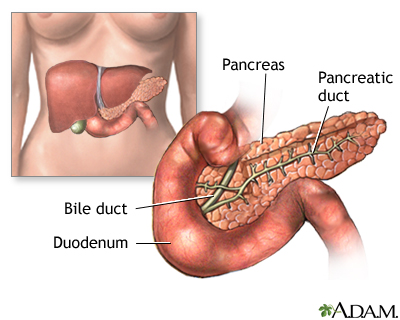
Pancreas

Pancreas

The pancreas is located behind the liver and is where the hormone insulin is produced. Insulin is used by the body to store and utilize glucose.

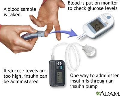
Insulin pump

Insulin pump

Various styles of insulin pumps may be utilized by people with diabetes to inject insulin into the body in a controlled, more convenient and discreet manner.

Glucose test

Glucose test

A person with diabetes constantly manages their blood's sugar (glucose) levels. After a blood sample is taken and tested, it is determined whether the glucose levels are low or high. If glucose levels are too low carbohydrates are ingested. If glucose in the blood is too high, the appropriate amount of insulin is administered into the body such as through an insulin pump.

Insulin pump

Insulin pump

The catheter at the end of the insulin pump is inserted through a needle into the abdominal fat of a person with diabetes. Dosage instructions are entered into the pump's small computer and the appropriate amount of insulin is then injected into the body in a calculated, controlled manner.

Type I diabetes

Type I diabetes

In response to high levels of glucose in the blood, the insulin-producing cells in the pancreas secrete the hormone insulin. Type I diabetes occurs when these cells are destroyed by the body’s own immune system.

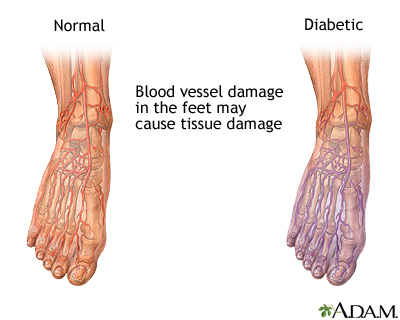
Diabetic blood circulation in foot

Diabetic blood circulation in foot

People with diabetes are at risk for blood vessel injury, which may be severe enough to cause tissue damage in the legs and feet.

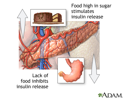
Food and insulin release

Food and insulin release

Insulin is a hormone secreted by the pancreas in response to increased glucose levels in the blood.

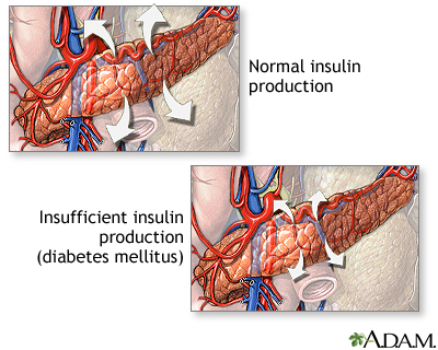
Insulin production and diabetes

Insulin production and diabetes

Insulin is a hormone produced by the pancreas that is necessary for cells to be able to use blood sugar.

Monitor blood glucose - series: Part 1


Part 1
Set up the meter according to the specific directions that come with your meter. Get the supplies ready, including a new test strip and disposable lancet. Place the lancet into the lancing device.

Definition

Diabetes is a life-long disease marked by high levels of sugar in the blood.

Causes, incidence, and risk factors

Diabetes can be caused by too little insulin (a hormone produced by the pancreas to control blood sugar), resistance to insulin, or both.

To understand diabetes, it is important to first understand the normal process of food metabolism. Several things happen when food is digested:

  • A sugar called glucose enters the bloodstream. Glucose is a source of fuel for the body.
  • An organ called the pancreas makes insulin. The role of insulin is to move glucose from the bloodstream into muscle, fat, and liver cells, where it can be used as fuel.

People with diabetes have high blood sugar. This is because their pancreas does not make enough insulin or their muscle, fat, and liver cells do not respond to insulin normally, or both.

There are three major types of diabetes:

  • Type 1 diabetes is usually diagnosed in childhood. The body makes little or no insulin, and daily injections of insulin are needed to sustain life.
  • Type 2 diabetes is far more common than type 1 and makes up most of all cases of diabetes. It usually occurs in adulthood. The pancreas does not make enough insulin to keep blood glucose levels normal, often because the body does not respond well to the insulin. Many people with type 2 diabetes do not know they have it, although it is a serious condition. Type 2 diabetes is becoming more common due to the growing number of older Americans, increasing obesity, and failure to exercise.
  • Gestational diabetes is high blood glucose that develops at any time during pregnancy in a woman who does not have diabetes.

Diabetes affects more than 20 million Americans. About 54 million Americans have prediabetes. There are many risk factors for diabetes, including:

  • A parent, brother, or sister with diabetes
  • Obesity
  • Age greater than 45 years
  • Some ethnic groups (particularly African Americans, Native Americans, Asians, Pacific Islanders, and Hispanic Americans)
  • Gestational diabetes or delivering a baby weighing more than 9 pounds
  • High blood pressure
  • High blood levels of triglycerides (a type of fat molecule)
  • High blood cholesterol level
  • Not getting enough exercise

The American Diabetes Association recommends that all adults over age 45 be screened for diabetes at least every 3 years. A person at high risk should be screened more often.

Symptoms

High blood levels of glucose can cause several problems, including frequent urination, excessive thirst, hunger, fatigue, weight loss, and blurry vision. However, because type 2 diabetes develops slowly, some people with high blood sugar experience no symptoms at all.

Symptoms of type 1 diabetes:

  • Increased thirst
  • Increased urination
  • Weight loss in spite of increased appetite
  • Fatigue
  • Nausea
  • Vomiting

Patients with type 1 diabetes usually develop symptoms over a short period of time, and the condition is often diagnosed in an emergency setting.

Symptoms of type 2 diabetes:

  • Increased thirst
  • Increased urination
  • Increased appetite
  • Fatigue
  • Blurred vision
  • Slow-healing infections
  • Impotence in men

Signs and tests

A urine analysis may be used to look for glucose and ketones from the breakdown of fat. However, a urine test alone does not diagnose diabetes. The following blood glucose tests are used to diagnose diabetes:

  • Fasting blood glucose level -- diabetes is diagnosed if higher than 126 mg/dL on two occasions. Levels between 100 and 126 mg/dl are referred to as impaired fasting glucose or pre-diabetes. These levels are considered to be risk factors for type 2 diabetes and its complications.
  • Random (non-fasting) blood glucose level -- diabetes is suspected if higher than 200 mg/dL and accompanied by the classic symptoms of increased thirst, urination, and fatigue. (This test must be confirmed with a fasting blood glucose test.)
  • Oral glucose tolerance test -- diabetes is diagnosed if glucose level is higher than 200 mg/dL after 2 hours (This test is used more for type 2 diabetes.)

You should also ask your doctor how often to you need your hemoglobin A1c (HbA1c) level checked. The HbA1c is a measure of average blood glucose during the previous 2 to 3 months. It is a very helpful way to determine how well treatment is working.

Ketone testing is another test that is used in type 1 diabetes. Ketones are produced by the breakdown of fat and muscle, and they are harmful at high levels. The ketone test is done using a urine sample. High levels of blood ketones may result in a serious condition called ketoacidosis. Ketone testing is usually done at the following times:

  • When the blood sugar is higher than 240 mg/dL
  • During acute illness (for example, pneumonia, heart attack, or stroke)
  • When nausea or vomiting occur
  • During pregnancy

Treatment

There is no cure for diabetes. Treatment involves medicines, diet, and exercise to control blood sugar and prevent symptoms and complications.

LEARN THESE SKILLS

Basic diabetes management skills will help prevent the need for emergency care. These skills include:

  • How to recognize and treat low blood sugar (hypoglycemia) and high blood sugar (hyperglycemia)
  • What to eat and when
  • How to take insulin or oral medication
  • How to test and record blood glucose
  • How to test urine for ketones (type 1 diabetes only)
  • How to adjust insulin or food intake when changing exercise and eating habits
  • How to handle sick days
  • Where to buy diabetes supplies and how to store them

After you learn the basics of diabetes care, learn how the disease can cause long-term health problems and the best ways to prevent these problems. People with diabetes need to review and update their knowledge, because new research and improved ways to treat diabetes are constantly being developed.

SELF-TESTING

If you have diabetes, your doctor may tell you to regularly check your blood sugar levels at home. There are a number of devices available, and they use only a drop of blood. Self-monitoring tells you how well diet, medication, and exercise are working together to control your diabetes and can help your doctor prevent complications.

The American Diabetes Association recommends that premeal blood sugar levels fall in the range of 80 to 120 mg/dL and bedtime blood levels fall in the range of 100 to 140 mg/dL. Your doctor may adjust this depending on your circumstances.

WHAT TO EAT

You should work closely with your health care provider to learn how much fat, protein, and carbohydrates you need in your diet. A registered dietician can be very helpful in planning dietary needs.

People with type 1 diabetes should eat at about the same times each day and try to be consistent with the types of food they choose. This helps to prevent blood sugars from becoming extremely high or low.

Persons with type 2 diabetes should follow a well-balanced and low-fat diet.

HOW TO TAKE MEDICATION

Medications to treat diabetes include insulin and glucose-lowering pills called oral hypoglycemic drugs.

Persons with type 1 diabetes cannot make their own insulin, so daily insulin injections are needed. Insulin does not come in pill form. Injections that are generally needed one to four times per day. Some people use an insulin pump, which is worn at all times and delivers a steady flow of insulin throughout the day. Other people may use a new type of inhaled insulin.

Insulin preparations differ in how quickly they start to work and how long they remain active. Sometimes different types of insulin are mixed together in a single injection. The types of insulin to use, the doses needed, and the number of daily injections are chosen by a health care professional trained to provide diabetes care.

People who need insulin are taught to give themselves injections by their health care providers or diabetes educators.

Unlike type 1 diabetes, type 2 diabetes may respond to treatment with exercise, diet, and medicines taken by mouth. There are several types of medicines used to lower blood glucose in type 2 diabetes. They fall into one of three groups:

  1. Medications called oral sulfonylureas that increase insulin production by the pancreas.
  2. Medications called thiazolidinediones that help increase the cell's sensitivity (responsiveness) to insulin.
  3. Medications that delay absorption of glucose from the gut. These include acarbose and miglitol.

There are some injectable medicines used to lower blood sugar. They include exenatide and pramlintide.

Most persons with type 2 diabetes will need more than one medication for good blood sugar control within 3 years of starting their first medication. Different groups of medications may be combined or used with insulin.

Some people with type 2 diabetes find they no longer need medication if they lose weight and increase activity, because when their ideal weight is reached, their own insulin and a careful diet can control their blood glucose levels.

It is unknown if hypoglycemic medicines taken by mouth are safe for use in pregnancy. Women who have type 2 diabetes and take these medications may be switched to insulin during pregnancy and while breastfeeding.

Gestational diabetes is treated with insulin and changes in diet.

EXERCISE

Regular exercise is especially important for people with diabetes. It helps with blood sugar control, weight loss, and high blood pressure. People with diabetes who exercise are less likely to experience a heart attack or stroke than diabetics who do not exercise regularly. You should be evaluated by your physician before starting an exercise program.

Here are some exercise considerations:

  • Choose an enjoyable physical activity that is appropriate for your current fitness level.
  • Exercise every day, and at the same time of day, if possible.
  • Monitor blood glucose levels before and after exercise.
  • Carry food that contains a fast-acting carbohydrate in case you become hypoglycemic during or after exercise.
  • Carry a diabetes identification card and a mobile phone or change for a payphone in case of emergency.
  • Drink extra fluids that do not contain sugar before, during, and after exercise.

Changes in exercise intensity or duration may need changes in diet or medication dose to keep blood sugar levels from going too high or low.

FOOT CARE

People with diabetes are prone to foot problems because of the likelihood of damage to blood vessels and nerves and a decreased ability to fight infection. Problems with blood flow and damage to nerves may cause an injury to the foot to go unnoticed until infection develops. Death of skin and other tissue can occur.

If left untreated, the affected foot may need to be amputated. Diabetes is the most common condition leading to amputations.

To prevent injury to the feet, people with diabetes should adopt a daily routine of checking and caring for the feet as follows:

  • Check your feet every day, and report sores or changes and signs of infection.
  • Wash your feet every day with lukewarm water and mild soap, and dry them thoroughly.
  • Soften dry skin with lotion or petroleum jelly.
  • Protect feet with comfortable, well-fitting shoes.
  • Exercise daily to promote good circulation.
  • See a podiatrist for foot problems or to have corns or calluses removed.
  • Remove shoes and socks during a visit to your health care provider and remind him or her to examine your feet.
  • Stop smoking, which hinders blood flow to the feet.

Expectations (prognosis)

With good blood glucose and blood pressure control, many of the complications of diabetes can be prevented.

Studies have shown that strict control of blood sugar and blood pressure levels in persons with diabetes helps reduce the risk of kidney disease, eye disease, nervous system disease, heart attack, and stroke.

Complications

Calling your health care provider

Go to the emergency room or call the local emergency number (such as 911) if symptoms of ketoacidosis occur:

  • Increased thirst and urination
  • Nausea
  • Deep and rapid breathing
  • Abdominal pain
  • Sweet-smelling breath
  • Loss of consciousness

Go to the emergency room or call the local emergency number if symptoms of extremely low blood sugar (hypoglycemic coma or severe insulin reaction) occur:

Prevention

Maintaining an ideal body weight and an active lifestyle may prevent the onset of type 2 diabetes. Currently there is no way to prevent type 1 diabetes.


Thursday, September 6, 2007

Aging Changes in The Heart and Blood Vessels

Taking your carotid pulse

Taking your carotid pulse

The carotid arteries take oxygenated blood from the heart to the brain. The pulse from the carotids may be felt on either side of the front of the neck just below the angle of the jaw. This rhythmic "beat" is caused by varying volumes of blood being pushed out of the heart toward the extremities.

Circulation of blood through the heart

Circulation of blood through the heart

The heart is a large muscular organ which constantly pushes oxygen-rich blood to the brain and extremities and transports oxygen-poor blood from the brain and extremities to the lungs to gain oxygen. Blood comes into the right atrium from the body, moves into the right ventricle and is pushed into the pulmonary arteries in the lungs. After picking up oxygen, the blood travels back to the heart through the pulmonary veins into the left atrium, to the left ventricle and out to the body's tissues through the aorta.

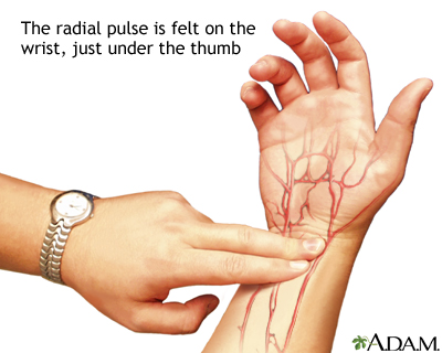
Radial pulse

Radial pulse

Arteries carry oxygenated blood away from the heart to the tissues of the body; veins carry blood depleted of oxygen from the same tissues back to the heart. The arteries are the vessels with the "pulse", a rhythmic pushing of the blood in the heart followed by a refilling of the heart chamber. To determine heart rate, one feels the beats at a pulse point like the inside of the wrist for 10 seconds, and multiplies this numbers by six. This is the per-minute total.

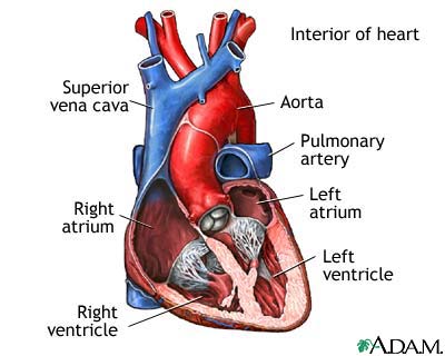
Normal heart anatomy (cut section)

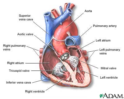
Normal heart anatomy (cut section)

The normal heart viewed so that major valves can be seen.

Effects of age on blood pressure

Effects of age on blood pressure

Blood vessels become less elastic with age. The "average" blood pressure increases from 120/70 to 150/90 and may persist slightly high even if treated. The blood vessels respond more slowly to a change in body position.

Information

Some changes in the heart and blood vessels normally occur with age, but many others are modifiable factors that, if not treated, can lead to heart disease.

BACKGROUND

The heart has two sides. The right side pumps blood to the lungs to receive oxygen and get rid of carbon dioxide. The left side pumps oxygen-rich blood to the body.

Blood flows out of the heart through arteries, which branch out and get smaller and smaller as they go into the tissues. In the tissues, they become tiny capillaries.

Capillaries are where the blood gives up oxygen and nutrients to the tissues, and receives carbon dioxide and wastes back from the tissues. Then, the vessels begin to collect together into larger and larger veins, which return blood to the heart.

Aging causes changes in the heart and in the blood vessels. Heart and blood vessel diseases are some of the most common disorders in the elderly.

AGING CHANGES

Heart

  • Normal changes in the heart include deposits of the "aging pigment," lipofuscin. The heart muscle cells degenerate slightly. The valves inside the heart, which control the direction of blood flow, thicken and become stiffer. A heart murmur caused by valve stiffness is fairly common in the elderly.
  • The heart has a natural pacemaker system that controls heartbeat. Some of the pathways of this system may develop fibrous tissue and fat deposits. The natural pacemaker (the SA node) loses some of its cells. These changes may result in a slightly slower heart rate.
  • Heart changes cause the ECG of a normal, healthy aged person to be slightly different than the ECG of a healthy younger adult. Abnormal rhythms (arrhythmias) such as atrial fibrillation are common in older people, which may be caused by heart disease.
  • A slight increase in the size of the heart, especially the left ventricle, is not uncommon. The heart wall thickens, so the amount of blood that the chamber can hold may actually decrease despite the increased overall heart size. The heart may fill more slowly.

Blood vessels

  • The main artery from the heart (aorta) becomes thicker, stiffer, and less flexible. This is probably related to changes in the connective tissue of the blood vessel wall. This makes the blood pressure higher and makes the heart work harder, which may lead to hypertrophy (thickening of the heart muscle). The other arteries also thicken and stiffen. In general, most elderly people experience a moderate increase in blood pressure.
  • Receptors, called baroreceptors, monitor the blood pressure and make changes to help maintain a fairly constant blood pressure when a person changes positions or activities. The baroreceptors become less sensitive with aging. This may explain the relatively common finding of orthostatic hypotension, a condition in which the blood pressure falls when a person goes from lying or sitting to standing, resulting in dizziness.
  • The wall of the capillaries thickens slightly. This may cause a slightly slower rate of exchange of nutrients and wastes.

Blood

  • The blood itself changes slightly with age. Aging causes a normal reduction in total body water. As part of this, there is less fluid in the bloodstream, so blood volume decreases.
  • The number of red blood cells (and correspondingly, the hemoglobin and hematocrit levels) are reduced. This contributes to fatigue. Most of the white blood cells stay at the same levels, although certain white blood cells important to immunity (lymphocytes) decrease in number and ability to fight off bacteria. This reduces the ability to resist infection.

EFFECT OF CHANGES

Under normal circumstances, the heart continues to adequately supply all parts of the body. However, an aging heart may be slightly less able to tolerate increased workloads, because changes reduce this extra pumping ability (reserve heart function).

Some of the things that can increase heart workload include illness, infections, emotional stress, injuries, extreme physical exertion, and certain medications.

COMMON PROBLEMS

  • Heart and blood vessel diseases are fairly common in older people. Common disorders include high blood pressure and orthostatic hypotension.
  • Arteriosclerosis (hardening of the arteries) is very common. Fatty plaque deposits inside the blood vessels cause it to narrow and can totally block blood vessels.
  • Coronary artery disease is fairly common.
  • Angina (chest pain caused by temporarily reduced blood flow to the heart muscle), shortness of breath with exertion and heart attack can result from coronary artery disease.
  • Abnormal heart rhythms (arrhythmias) of various types can occur.
  • Heart failure is also very common in the elderly. In people older than 75, heart failure occurs 10 times more often than in younger adults.
  • Valve diseases are fairly common. Aortic stenosis, or narrowing of the aortic valve, is the most common valve disease in the elderly.
  • Anemia may occur, possibly related to malnutrition, chronic infections, blood loss from the gastrointestinal tract, or as a complication of other diseases or medications.
  • Transient ischemic attacks (TIA) or strokes can occur if blood flow to the brain is disrupted.

Other problems with the heart and blood vessels include the following:

PREVENTION

You can help your circulatory system (heart and blood vessels). Heart disease risk factors that you have some control over include high blood pressure, cholesterol levels, diabetes, obesity, and smoking.

  • Eat a heart-healthy diet with reduced amounts of saturated fat and cholesterol, and control your weight. Follow your health care provider's recommendations for treatment of high blood pressure, high cholesterol or diabetes. Minimize or stop smoking.
  • Moderate exercise is one of the best things you can do to keep your heart, and the rest of your body, healthy. Consult with your health care provider before beginning a new exercise program. Exercise moderately and within your capabilities, but do it regularly.
  • People who exercise usually have less body fat and smoke less than people who do not exercise. They also tend to have fewer blood pressure problems and less heart disease.
  • Exercise may help prevent obesity and helps people with diabetes control their blood sugar.
  • Exercise may help you maintain your maximum abilities as much as possible and reduces stress.

Heart MRI

MRI scans

MRI scans

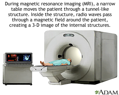
MRI stands for magnetic resonance imaging. It allows imaging of the interior of the body without using x-rays or other types of ionizing radiation. An MRI scan is capable of showing fine detail of different tissues.

Definition

Heart magnetic resonance imaging (MRI) is a method that uses powerful magnets and radio waves to create pictures of the heart. It does not use radiation (x-rays).

The test may be done as part of a chest MRI.

How the test is performed

You will be asked to lie on a narrow table, which slides into a large tunnel-like tube. The health care provider may inject a dye through one of your veins. This helps certain diseases and organs show up better on the images.

Unlike and computed tomographic (CT) scans, MRI does not use radiation. Instead, it uses powerful magnets and radio waves. The magnetic field produced by an MRI forces certain atoms in your body to line up in a certain way. It's similar to how the needle on a compass moves when you hold it near a magnet.

The radio waves are sent toward these atoms and bounce back, and a computer records the signal. Different types of tissues send back different signals. For example, healthy tissue sends back a slightly different signal than cancerous tissue.

A technologist will operate the machine from a room next door and watch you during the entire study.

Several sets of images are usually needed. Each one takes about 2-15 minutes. A complete scan may take up to 1 hour. Newer scanners may complete the process in less time.

How to prepare for the test

There is usually no preparation needed. An MRI can be done immediately after other imaging studies. If contrast (dye) or sedation is used, you may be asked not to eat for 4 to 6 hours prior to the scan.

The strong magnetic fields created during an MRI can interfere with certain implants, particularly cardiac pacemakers. People with cardiac pacemakers can not have an MRI and should not enter the MRI area.

If you have any of the following metallic objects in your body, you should not get an MRI:

  • Brain aneurysm clips
  • Certain artificial heart valves
  • Inner ear (cochlear) implants
  • Older vascular stents
  • Recently placed artificial joints

You will be asked to sign a consent form that says you do not have any of these items in your body. You may be asked to wear a hospital gown.

Certain metallic objects are not allowed into the room.

  • Items such as jewelry, watches, credit cards, and hearing aids can be damaged.
  • Pins, hairpins, metal zippers, and similar metallic items can distort the images.
  • Removable dental work should be taken out just prior to the scan.

When the MRI magnet is turned on, pens, pocketknives, and eyeglasses may fly across the room. This can be dangerous, so such items are not allowed into the scanner area.

How the test will feel

A heart MRI exam causes no pain. Some people may become anxious when inside the scanner. If you have difficulty lying still or are very anxious, you may be given a mild sedative. Excessive movement can blur MRI images and cause errors.

The table may be hard or cold, but you can request a blanket or pillow. The machine produces loud thumping and humming noises when turned on. Ear plugs are usually given to help reduce the noise.

An intercom in the scanner allows you to speak to the person operating the exam at any time. Some MRIs have televisions and special headphones that you can use to help the time pass.

There is no recovery time, unless sedation was necessary. (You will need someone to drive you home if sedation was given.) After an MRI scan, you can resume your normal diet, activity, and medications, unless otherwise instructed by your doctor.

Why the test is performed

MRI provides detailed pictures of the heart and blood vessels from many views.

It may be used to diagnose:

  • Heart muscle damage after a heart attack
  • Birth defects of the heart
  • Heart tumors and growths

MRI is sometimes used to avoid the dangers of angiography, repeated exposure to radiation, or the use of iodine-based dye (contrast).

It may provide additional information when an echocardiogram is unclear.

What abnormal results mean

The sensitivity of MRI depends, in part, on the experience of the radiologist.

A heart MRI may reveal the following disorders:

What the risks are

There is no ionizing radiation involved in MRI, and there have been no documented significant side effects of the magnetic fields and radio waves used on the human body to date.

The most common type of contrast (dye) used is gadolinium. It is very safe. Allergic reactions to the substance rarely occur. The person operating the machine will monitor your heart rate and breathing as needed.

People have been harmed in MRI machines when they did not remove metal objects from their clothes or when metal objects were left in the room by others.

MRI is usually not recommended for traumatic injuries, because traction and life-support equipment cannot safely enter the scanner area, and scans can take a long time.

Special considerations

MRI is more accurate than CT scan or other tests for certain conditions, but less accurate for others. Disadvantages include the high cost, long duration of the scan, and sensitivity to movement. People with claustrophobia or who are confused or anxious may have difficulty lying still for the relatively long scan times. MRI is not portable and is incompatible with some metallic implants, life support devices, traction, apparatus, and similar equipment.

MRI is superior in most cases in which differentiation of soft tissues is necessary. It can view organs without obstruction by bone and foreign bodies. It is capable of showing the tissues from multiple viewpoints and is a non invasive way to evaluate blood flow.

Heart Disease and Women

Heart, section through the middle

Heart, section through the middle

The interior of the heart is composed of valves, chambers, and associated vessels.

Acute MI

Acute MI

A heart attack or acute myocardial infarction (MI) occurs when one of the arteries that supplies the heart muscle becomes blocked. Blockage may be caused by spasm of the artery or by atherosclerosis with acute clot formation. The blockage results in damaged tissue and a permanent loss of contraction of this portion of the heart muscle.

Information

Mention the term "heart attack" and most people imagine a pudgy, middle-aged man drenched in sweat and clutching his chest. Few people seem to consider cardiovascular disease (CVD) as a woman's disease.

But according to the American Heart Association, cardiovascular disease is the leading killer of women over age 25. It kills nearly twice as many women in the United States than all types of cancer, including breast cancer. Only 13 percent of women think heart disease is a threat to their health.

The misleading notion that heart disease is not a real problem for women can be blamed in part on medical research. For a very long time, heart disease studies have focused primarily on men. Changes are under way, but some doctors still fail to recognize the warning signs displayed by female patients.

EARLY HEART SIGNS

Studies have shown that women have undiagnosed warning signs weeks, months, and even years before having a heart attack.

Significant differences exist in the symptoms displayed by women and men. Men typically experience the "classic" heart attack signs: tightness in the chest, arm pain, and shortness of breath. Women's symptoms -- nausea, an overwhelming fatigue, and dizziness -- are strikingly different and are often chalked up to stress. Women have reported that they have had a hard time getting their doctors to listen to them about these early warning symptoms.

Unusual fatigue, trouble sleeping, shortness of breath, indigestion, and anxiety were the top 5 symptoms reported by both black and white women in the study. However, black women had more intense episodes and reported them more often.

ACT IN TIME

Recognizing and treating a heart attack right away dramatically improves a patient's chance for survival. The typical American, however, waits 2 hours before calling for help.

Studies have shown that drugs that dissolve coronary blood clots during a heart attack can reduce the death rate in both men and women, although women have a higher risk of stroke from the therapy. Unfortunately, statistics show that a woman in the midst of having a heart attack receives clot-busting therapy much later than a man would.

Women coming into the hospital for a heart attack have a higher death rate and higher risk of complications. A premenopausal woman having a heart attack has twice the death rate of a similarly aged man, according to experts.

Know the warning signs and always call 911 within 5 minutes of the onset of symptoms. By acting quickly, a heart attack victim is less likely to experience cardiac arrest (where the heart stops beating).

PREVENTION TIED TO BELIEF

There is no denying that an ounce of prevention is worth a pound of cure. But preventing a disease means believing you are actually at risk -- and many women fail to see that.

Women are advised to take charge of their health by working with their doctor to address risk factors, and keep tabs on cholesterol levels, blood pressure, and lifestyle.

According to the American Heart Association, low blood levels of "good" cholesterol (high density lipoprotein, or HDL) are a stronger predictor of heart disease death in women than in men. Also, a major study showed that post-menopausal women taking hormone replacement therapy have an increased risk of heart attack and death by coronary artery disease.

WOMEN & HEART DISEASE

  • Cardiovascular disease kills about one woman a minute.
  • Sixty-four percent of women who die suddenly of heart disease have no previous symptoms.
  • More women than men will die within the first year after a heart attack
  • The rates of women who die from cardiovascular disease are much higher than those who die from breast cancer.

Source: American Heart Association/Go Red For Women

The American Heart Association has published guidelines regarding prevention of cardiovascular disease in women. Women are categorized based on their likelihood of experiencing a cardiovascular event (heart attack, stroke, death) in the next 10 years:

  • High risk -- the woman has a greater than 20% chance of an event in next 10 years. Examples of women at high risk include those with have a history of stroke, heart disease, vascular disease in legs, abdominal aortic aneurysm (AAA), diabetes, and chronic kidney disease.
  • Intermediate risk -- the woman has a 10 - 20% chance of event in next 10 years. Examples of women at intermediate risk include those with calcium in coronary arteries, metabolic syndrome, multiple heart risk factors, and women with family history of early heart disease.
  • Lower risk -- the woman has less than a 10% chance of event in next 10 years. Women at the lowest risk have none of the above-mentioned conditions and one or less risk factors for heart disease.

The guidelines recommend the following lifestyle changes:

  • Do not smoke or use tobacco.
  • Maintain a healthy weight. Women who need to lose or keep off weight should get at least 60 - 90 minutes of moderate-intensity exercise on most days. To maintain your weight, get at least 30 minutes of exercise a day, preferably at least 5 days a week.
  • Women who recently had a heart attack, angina, angioplasty, or a stent procedure should join a cardiac rehabilitation program.
  • Eat a heart healthy diet. The diet includes a variety of fruits, vegetables, grains, low-fat or nonfat dairy products, fish, legumes, and sources of protein low in saturated fat.
  • Watch your weight. Women should strive for a body mass index (BMI) between 18.5 and 24.9 and a waist smaller than 35 inches.
  • Get checked and treatment, if necessary, for depression.
  • High-risk women should take omega-3 fatty acids supplements.
  • Folic acid supplements may be considered in some high-risk women if a higher than normal level of homocysteine has been detected. (These should not be taken after coronary revascularization.)

TREATMENT RECOMMENDATIONS FOR WOMEN

Keep blood pressure under 120/80 mm Hg. Blood pressure drugs should be used when blood pressure is higher than 140/90 mm Hg. (Persons with diabetes may need medication at lower levels.)

Keep cholesterol levels under control:

  • LDL should be under 100 mg/dL
  • HDL should be greater than 50 mg/dL
  • Triglycerides should be less than 150 mg/dL

Depending on a woman's level of risk (low, intermediate, high), dietary changes and medicines may be needed to control cholesterol levels.

New guidelines no longer recommend hormone replacement therapy, antioxidant supplements, or folic acid to prevent heart disease in women.

Aspirin therapy (dose 75 mg to 325 mg a day) or a drug called clopidogrel may be prescribed for women at high risk for heart disease. Aspirin therapy is recommended for women over age 65 to prevent heart attack and stroke as long as blood pressure is controlled and the benefit is likely to outweigh the risk of gastrointestinal side effects. Regular use of aspirin is not recommended for healthy women under age 65 to prevent heart attacks.

Beta blockers should be used in all women with a history of heart attack or who have chronic heart disease, unless there is a medical reason not to.

ACE inhibitors should be used in high-risk women unless there is a medical reason not to.

Angiotensin receptor blockers should be used in women with heart failure or an ejection fraction less than 40% in whom ACE inhibitors did not work.

See also:

Heart Disease and Vitamin E

Vitamin E and heart disease

Vitamin E and heart disease

There is no conclusive evidence that taking vitamin E supplements can reduce your risk of heart disease. In fact, taking 400 IU of vitamin E (or higher) may actually be harmful.

Information

Antioxidants such as vitamin E (also called tocopherol) protect cells in the body from oxidation. Oxidation is a process that leads to cell damage. It may play an important role in atherosclerosis -- the development of plaque in blood vessels that can cause heart disease and stroke.

Eating foods rich in antioxidants (like vitamin E and vitamin C, carotenoids, and selenium) may lower your risk of heart disease. Such foods include fruits, vegetables, whole grains, nuts, and seeds. According to studies, however, taking extra anti-oxidant pills is probably NOT beneficial.

The current recommendation by the American Heart Association is to make sure you include these important nutrients in your diet, but not to take supplements. Because foods rich in vitamin E and selenium are high in fat, you may want to work with a dietitian to find the best food sources for you.

Heart Disease and Diet

Healthy diet

Healthy diet

For a healthy diet, replace unhealthy and fattening foods with healthier alternatives, such as fresh fruits and vegetables.

Fish in diet

Fish in diet

Fish is a great alternative to red meat. Fish is a healthy, lean protein and contains a type of fat called "omega-3," which may help protect the heart.

Fruits and vegetables

Fruits and vegetables

A healthy diet includes adding vegetables and fruit every day. Vegetables like broccoli, green beans, leafy greens, zucchini, cauliflower, cabbage, carrots, and tomatoes are low in calories and high in fiber, vitamins, and minerals. Many studies have shown that eating plenty of vegetables is extremely healthy. Try to eat about 3 to 5 servings every day. Fruit is also a good source of fiber, vitamins, and minerals. You should try to eat about 2 to 3 servings of fruit each day.

Definition

A healthy diet is a major factor in reducing your risk of heart disease.

Function

A healthy diet and lifestyle can reduce your risk of:

Food Sources

Most fruits and vegetables are appropriate for a heart-healthy diet. They are good sources of fiber, vitamins, and minerals. Most are low in fat, calories, sodium, and cholesterol.

Dairy products and milk are good sources of protein, calcium, the B vitamins niacin and riboflavin, and the vitamins A and D. Use skim, 1/2%, or 1% milk. Cheese, yogurt, and buttermilk should be low-fat or nonfat.

Eat low-fat breads, cereals, crackers, rice, pasta, and starchy vegetables (like peas, potatoes, corn, winter squash, and lima beans). These foods are high in the B vitamins, iron, and fiber. At the same time, they are low in fat and cholesterol.

Avoid baked goods such as butter rolls, cheese crackers, and croissants, cream sauces for pasta and vegetables, and cream soups.

Meat, poultry, seafood, dried peas, lentils, nuts, and eggs are good sources of protein, the B vitamins, iron, and other vitamins and minerals.

  • Eat skinless poultry, very lean beef, lamb, veal, and pork, lentils, legumes, dried beans and peas, egg whites, and wild game.
  • Avoid duck, goose, marbled meats (such as a ribeye steak), prime cuts of high-fat meats, organ meats such as kidneys and liver, and prepared meats such as sausage, frankfurters, and high-fat lunch meats.

Limit oils and fats. They are high in fat and calories, and people should eat less of all types of fat. Some fats are better choices than others but should still be used in moderate amounts.

  • Use liquid vegetable oils such as safflower, soybean, corn, sesame, olive, canola, avocado, and cottonseed. Use margarines made from any of these oils in their tub or squeeze form, not their stick form. Salad dressings and mayonnaise should be made with the recommended oils.
  • Seeds, nuts, olives, avocados, and peanut butter are also acceptable in moderate amounts.
  • Avoid butter, lard, bacon, shortening, sour cream, whipping cream, and coconut, palm, or palm kernel oil. These contain saturated fats and are not recommended.

Diet recommendations for children over the age of 2 years are similar to those of adults. Children and teenagers must have enough calories to support growth and activity level while they achieve and maintain a desirable body weight.

Children following low-fat diets may have difficulty maintaining desired levels of growth. Consult a physician or dietitian for assistance in planning adequate low-fat meals for children and adolescents.

A consultation with a registered dietitian is helpful. The American Heart Association has local chapters in every state. They are an excellent resource for information on heart disease.

Recommendations

  • Maintain your ideal body weight and balance the number of calories you eat with the number you use each day. You can ask a dietician or a health care professional to help you determine these numbers.
  • Limit your intake of foods high in calories or low in nutrition, including foods like soft drinks and candy that have lots of sugars.
  • Eat five or more servings per day of fruits and vegetables.
  • Eat six or more servings per day of grain products, including whole grains. Grain products provide fiber, vitamins, minerals, and complex carbohydrates. The daily calories should be appropriate for the maintenance of desirable body weight and should support growth in children and adolescents.
  • Reduce total fat intake. Limit foods high in saturated fat, trans fat, cholesterol, and partially hydrogenated oils. Reduce or avoid saturated fat when possible. Saturate fat raises your cholesterol level. Choose liquid or tub margarine, canola oil, or olive oil. These have 2g or less of saturated fat per serving.
  • Eat less than 300 mg of dietary cholesterol daily. (For example, one egg yolk contains an average of 213 mg.)
  • Limit the amount of salt (sodium chloride) you eat. You should eat less than 2,400 mg of salt per day. Check food labels, since many foods contain salt.
  • Exercise regularly. For example, walk for at least 30 minutes a day.
  • Limit the amount of alcohol you drink. Women should have no more than one alcoholic drink (such as red wine) per day. Men should not drink more than two. While major studies have linked some alcohol consumption to health benefits, excessive drinking can do more harm than good.

EATING TIPS

  • To reduce fat and cholesterol, eat no more than 6 cooked ounces of meat, poultry, and fish daily. One serving of meat should be about the size of a deck of cards on your plate.
  • Use skinless turkey, chicken, fish, or lean red meat to reduce the amount of saturated fat in your diet. Lean, 3 ounce cuts of red meat may be used occasionally.
  • Trim all the visible fat prior to cooking the meat. Eat two servings of fish per week. Cook by baking, broiling, roasting, steaming, boiling, or microwaving rather than deep fat frying. For the main entree, use less meat or have meatless meals a few times a week. Use smaller amounts of meat to reduce the total fat content of the meal. Use no more than 5-8 teaspoons of fats or oils per day for salads, cooking, and baking.
  • To reduce high cholesterol, do not use more than 3-4 egg yolks per week, including eggs used in cooking. Eat less organ meat (such as liver) and shellfish (such as shrimp and lobster).
  • To reduce salt, reduce the amount of table salt used, and limit the use of prepared foods that have salt added to them, such as canned soups and vegetables, cured meats, and some frozen meals. Always check the nutrition label for the sodium content per serving.

Heart Bypass Surgery

Heart, front view
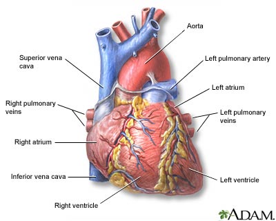
Heart, front view

The external structures of the heart include the ventricles, atria, arteries and veins. Arteries carry blood away from the heart while veins carry blood into the heart. The vessels colored blue indicate the transport of blood with relatively low content of oxygen and high content of carbon dioxide. The vessels colored red indicate the transport of blood with relatively high content of oxygen and low content of carbon dioxide.

Posterior heart arteries

Posterior heart arteries

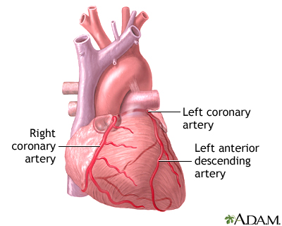
The coronary arteries supply blood to the heart muscle. The right coronary artery supplies both the left and the right heart; the left coronary artery supplies the left heart.

Anterior heart arteries

Anterior heart arteries

The coronary arteries supply blood to the heart muscle. The right coronary artery supplies both the left and the right heart; the left coronary artery supplies the left heart.

Heart bypass surgery - series: Normal anatomy

Normal anatomy
The heart muscle is supplied blood through the coronary arteries. The left coronary artery supplies blood to the left ventricle. The right coronary artery supplies blood to the right ventricle.

Description

Coronary arteries are the small blood vessels that supply the heart muscle with oxygen and nutrients. Fats and cholesterol can accumulate inside these small arteries, and the arteries can gradually become clogged. This buildup of fat and cholesterol plaque is called atherosclerosis.

When one or more of the coronary arteries becomes partially or totally blocked, the heart does not get an adequate blood supply. This is called ischemic heart disease or coronary artery disease (CAD). It can cause chest pain (angina).

Sometimes CAD does not cause pain until the blood supply to the heart becomes critically low, and the muscle begins to die. The first symptom of CAD in this case may be a potentially deadly heart attack. Symptomless CAD is especially common in diabetics.

OVERVIEW OF THE PROCEDURE

Heart bypass surgery creates a detour or "bypass" around the blocked part of a coronary artery to restore the blood supply to the heart muscle. The surgery is commonly called Coronary Artery Bypass Graft, or CABG (pronounced "cabbage").

After the patient is anesthetized and completely free from pain, the heart surgeon makes an incision in the middle of the chest and separates the breastbone.

Through this incision, the surgeon can see the heart and aorta (the main blood vessel leading from the heart to the rest of the body). After surgery, the breastbone will be rejoined with wire and the incision will be sewn closed.

ARTERY AND VEIN GRAFTS

If a vein from the leg, called the saphenous vein, is to be used for the bypass, an incision is made in the leg and the vein removed. The vein is located on the inside of the leg, running from the ankle to the groin. The saphenous vein normally does only about 10% of the work of circulating blood from the leg back to the heart. Therefore, it can be taken out without harming the patient or harming the leg.

It is common for the leg to swell slightly during recovery from the surgery, but this is only temporary and is treated by elevating the leg.

The internal mammary artery (IMA) can also be used as the graft. This has the advantage of staying open for many more years than the vein grafts, but there are some situations in which it cannot be used.

The left IMA, or LIMA, is an artery that runs next to the sternum on the inside of the chest wall. It can be disconnected from the chest wall without affecting the blood supply to the chest. It is commonly connected to the artery on the heart that supplies most of the muscle, the left anterior descending artery, or LAD.

Other arteries are also now being used in bypass surgery. The most common of these is the radial artery. This is one of the two arteries that supply the hand with blood. It can usually be removed from the arm without any impairment of blood supply to the hand.

TRADITIONAL APPROACH

In the traditional surgery, the patient is connected to the heart-lung machine, or bypass pump, which adds oxygen to the blood and circulates blood to other parts of the body during the surgery. This is necessary because the heart muscle must be stopped before the graft can be done.

One end of the graft is stitched to an opening below the blockage in the coronary artery. If the grafted vessel is the saphenous vein or the radial artery, its other end is stitched to an opening made in the aorta. If the grafted vessel is the mammary artery, its other end is already connected to the aorta.

The entire surgery can take 4-6 hours. After the surgery, the patient is taken to the Intensive Care Unit. For a few days after the surgery, the patient is connected to monitors and tubes.

OTHER TECHNIQUES

Other surgical techniques for this procedure are being used more frequently. One popular method is to avoid the use of the heart-lung machine. This is called off-pump coronary artery bypass or OPCAB. This operation allows the bypass to be created while the heart is still beating.

The advantage here is that use of the heart-lung machine can lead to some loss of memory and mental clarity, while with OPCAB, that risk is reduced because the heart isn't stopped, and the blood isn't oxygenated externally.

Another alternative is the use of smaller incisions that avoid splitting the breastbone. This is referred to as Minimally Invasive Direct Coronary Artery Bypass or MIDCAB.

Coronary bypass surgery can now be performed with the aid of a robot, which allows the surgeon to perform the operation without even being in the same room as the patient.

Indications

Coronary artery bypass surgery is a treatment option for ischemic heart disease (too little blood reaching the heart muscle). Coronary surgery is recommended when there is disease of the left main coronary artery, disease of three or more vessels (triple vessel disease), or nonsurgical management hasn't worked. Nonsurgical management includes medication and/or angioplasty.

The earliest symptoms of ischemic heart disease include angina (chest pain) and shortness of breath. A person may have no symptoms; have mild, intermittent chest pain; or have more pronounced and steady pain. Still others have CAD that is severe enough to make everyday activities difficult.

Symptoms that usually bring a person to a doctor are a feeling of heaviness, tightness, pain, burning, pressure, or squeezing. This is usually behind the breastbone, but sometimes it is also in the arms, neck, or jaw. Some people have heart attacks without ever having any of these symptoms first.

In cases where there are no symptoms, a doctor may suspect CAD and perform a stress test to determine if it is present. CAD is sometimes suspected if there is a family history of heart disease and a combination of other factors, including high blood cholesterol, diabetes, high blood pressure, cigarette smoking, and being male.

Because CAD varies so much from one person to another, the way it is diagnosed and treated will also vary. Heart bypass surgery is just one treatment.

Risks

When considering the risks of CABG, it is important to remember that bypass surgery has been performed for more than 30 years. Cardiovascular surgeons have received extensive training in bypass techniques.

It is the most frequently performed major surgery in the United States, with over a half million done each year. As with any surgery, the health of the patient prior to surgery is a major consideration in determining risks.

Health conditions that should be considered prior to surgery are:

  • Age -- patients over 70 are at a slightly higher risk for complications
  • Gender -- women have a slightly higher risk
  • Previous heart surgery -- puts a person at a higher risk
  • Having another serious medical condition (such as diabetes, peripheral vascular disease, kidney disease, or lung disease)

Possible risks in having CABG are:

  • Heart attack, which occurs in 5% of these surgeries
  • Stroke, which occurs in 5% of these surgeries (the risk is greatest in those over 70)
  • Blood clots
  • Death, which occurs in 1 - 2% of those who have the surgery (that means 95 - 98% have no serious complications)
  • Sternal wound infection, which occurs in 1 - 4% of these surgeries (this complication is most often associated with obesity, diabetes, or having had previous CABG)

In about 30% of patients, "post-pericardiotomy syndrome" can occur anywhere from a few days to 6 months after surgery. The symptoms of this syndrome are fever and chest pain. It can be treated with medication.

The incision in the chest or the graft site (if the graft was from the leg or arm) can be itchy, sore, numb, or bruised.

Some people report memory loss and loss of mental clarity or "fuzzy thinking" following CABG.

As with all surgeries, there is a risk for heavy bleeding. In case a transfusion is needed during or after surgery, ask your doctor about making arrangements for an "autologous" pre-operative blood donation (banking your own blood for surgery).

You may also have family or friends with a compatible blood type donate blood for your surgery. The hospital, Red Cross, or local blood bank can provide family members and friends with necessary information about blood donation for your surgery.

There are general risks from anesthesia. These include reactions to medications and problems breathing.

Expectations after surgery

Every year over one half million Americans have coronary bypass surgery to relieve symptoms and prolong their lives. In the majority of people who have the surgery, the grafts remain open and functioning for 10 to 15 years.

CABG will improve blood flow to the heart but NOT prevent the eventual recurrence of coronary blockage. Lifestyle changes are necessary -- such as not smoking, improved diet, regular exercise, and treating high blood pressure and high cholesterol.

Convalescence

After the operation, the patient will spend 5 - 7 days in the hospital, with the first 2 hours in an intensive-care unit (ICU). In the ICU, heart function is monitored continuously.

Patients may require the temporary assistance of a breathing tube for a few hours after surgery. Two to three tubes in the chest drain fluid from around the heart and are usually removed one to three days after surgery.

A urinary catheter in the bladder drains urine until the patient is able to void on his own. Intravenous lines (IV) provide fluids and medications. Nurses watch the monitors and check vital signs (pulse, temperature, breathing) constantly.

When constant monitoring is no longer needed, usually within 12 - 24 hours, the patient is moved to a regular or a transitional care unit. Activity is gradually resumed and the patient may begin a cardiac rehabilitation program within a few days. The incision in the chest does not bother most people after the first 48 - 72 hours.

After surgery, it takes 4 - 6 weeks to start feeling better. During recovery it is normal to:

  • Have a poor appetite -- it will take several weeks for it to return.
  • Have swelling in the leg if the graft was taken from the leg. Elevating the leg and wearing elastic TED hose for several weeks helps reduce swelling.
  • Have difficulty sleeping at night -- this will improve.
  • Have constipation.
  • Have mood swings and feel depressed -- this will get better.
  • Have difficulty with short-term memory or feel confused -- this also improves.

The full benefits from the operation may not be determined until 3 - 6 months after surgery. Sexual activities may be resumed 4 weeks after surgery. All activities that do not cause fatigue are permitted, and the schedule for resuming normal activities is determined with the physician.